
Should we be restricting access to healthcare?
September 18, 2014
Psychiatry in Malta – A look at Service Demand
September 23, 2014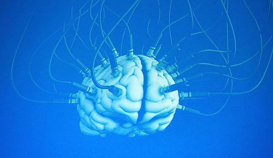
Transcranial Magnetic Stimulation... A Brief History
Faraday was the first to discover the principle of mutual induction in 1831 and in his law, quantified the conversion of electrical energy into magnetic fields and vice versa. Magnetic stimulation was first used towards the end of the 19th century in experiments conducted by d'Arsonval in 1896, the inventor of the galvanometer. Given the technical limitations of the day, d'Arsonval could only stimulate his subject’s retinas, eliciting a phenomenon known as magneto phosphenes, which is characterised by the experience of seeing light, when in fact; light had not penetrated the eye. It was not for over half a century from the pioneering works of d'Arsonval that the magnetic method was applied to neuromuscular stimulation. In 1965, Bickford and Fremming used an underpowered 500 Hz sinusoidal magnetic field in order to demonstrate muscular stimulation both in animals as well as in humans. This was subsequently replicated by Öberg in 1973. In 1982, Polson and his colleagues were the first to successfully stimulate superficial nerves via magnetic fields. However, it was not until the end of the 20 century that a device, the first transcranial magnetic stimulation device, capable of producing cortical depolarisation via magnetic stimulation, was created (Barker, Jalinous, & Freeston, 1985) and subsequently used for clinical experimentation, mainly by neurologists, for mapping of the human cortex. The first reported use of transcranial magnetic stimulation on psychiatric patients in an open study was in 1993 by Hofflich and his colleagues (1993), whose case report involving two treatment resistant patients, reported modest antidepressant effect. However, the early studies of the use of transcranial magnetic stimulation in the treatment of depression were limited by a number of factors. Primarily among these was the low-frequency stimulation that was employed and imprecise coil placement, as scientific knowledge to the importance of prefrontal coil placement was lacking.
Nomenclature
Transcranial Magnetic Stimulation can take the form of single-pulse, paired-pulse or repetitive Transcranial Magnetic Stimulation. Each application has different uses, from the mapping of motor cortical outputs and studying of brain behaviour relationship (single pulse) to the study of corticocortical interactions (paired pulse). Repetitive pulses delivered in ‘trains’, is known as repetitive Transcranial Magnetic Stimulation (rTMS), a term which has now been universally accepted as replacing earlier terms such as rapid-rate TMS or rapid TMS and refers to single Transcranial Magnetic Stimulation pulses which are regularly applied. In further sub-classification, the frequency of the pulses is used to differentiate between ‘fast’ or ‘high’ frequency rTMS (rates of more than 1 Hz) and ‘slow’ or ‘low’ frequency rTMS (rates of less than 1 Hz). This differentiation has been defined secondary to criteria, including varying physiological effects and degrees of related risk. A further subdivision known as patterned rTMS refers to short rTMS which is applied “in bursts at a high inner frequency interleaved by short pauses of no stimulation”, in protocols known as continuous (cTBS) or intermittent (iTBS) Theta-Burst Transcranial Magnetic Stimulation. However, the technical protocols go beyond the scope of this post.
Basic Principles
Transcranial Magnetic Stimulation uses the principle of inductance in order for electrical energy to cross the scalp and skull in order to do avoid the pain of direct percutaneous electrical stimulation (Wassermann, 1998):
“Transcranial Magnetic Stimulation involves placing a small coil of wire on the scalp and passing a powerful and rapidly changing current through it. This produces a magnetic field that passes unimpeded and relatively painlessly through the tissues of the head. The peak strength of the magnetic field is related to the magnitude of the current and the number of turns of wire in the coil. The magnetic field, in turn, induces a much weaker electrical current in the brain. The strength of the induced current is a function of the rate of change of the magnetic field, which is determined by the rate of change of the current in the coil. In order to produce enough current to excite neurons in the brain, the current passed through the coil must change within a few hundred microseconds.”
The power of the stimulators and coils is measured in Tesla which is the International System of Units of magnetic-field strength or magnetic-flux density, commonly denoted as T, defined in 1960 in honour of the inventor of the Tesla Coil, Nikola Tesla. Modern devices develop 1.5 - 4 tesla (T) measured at the coil’s surface with studies showing that cortical neurons are activated beneath the scalp to a depth of 1.5 – 2 cm, which effectively means that Transcranial Magnetic Stimulation with contemporary equipment does not appear to penetrate deeper than the cortex. Despite this, it has been shown that Transcranial Magnetic Stimulation has an effect on cells trans-synaptically beyond the site of stimulation, as evidenced by positron emission tomography studies, which show effects on distant cortical and subcortical sites (Wassermann, 1997).
As previously mentioned, Transcranial Magnetic Stimulation involves the administration on the scalp (in the primary motor cortex area) of a short (~ 200 ms) and powerful (0.2 to 4.0 Tesla) magnetic pulse through a coil, equal to 110-120% of the resting motor threshold (RMT), resulting in progressive increases in the amplitude of Motor Evoked Potentials (MEPs) and duration of the Cortical Silent Period (CSP). The magnetic pulse induces an electric current below the coil, over the cortical surface. This causes depolarisation of cell membranes as well as trans-synaptic depolarisation of cortical neurons. Originally, Transcranial Magnetic Stimulation was used in the field of clinical neurophysiology, for the functional evaluation of the cortico-spinal tract (Pellicciari, 2009). The procedure initially involved the administration of a single magnetic pulse, but by the mid-90's, technological advances allowed for the development of machines capable of delivering streams of repetitive magnetic pulses in rapid succession, with a frequency that could reach 100 Hz. This type of stimulation was called repetitive Transcranial Magnetic Stimulation and it was shown to be superior in its cortical interaction compared to single-pulse Transcranial Magnetic Stimulation.
Furthermore, repetitive Transcranial Magnetic Stimulation can be applied at low and/or at high frequency and, depending on the mode of application, may have different effects on cortical activity (Speer, 2000). The cut-off between high and low frequency is established empirically, on the basis of converging evidence indicating that repetitive Transcranial Magnetic Stimulation at less than 1 Hz reduces cortical excitability both locally as well as in functionally related areas, while repetitive Transcranial Magnetic Stimulation given at a frequency above 5 Hz, seems to have the opposite effect (Veniero, 2011). Therefore, it would seem that the modulation of neural excitability induced by repetitive Transcranial Magnetic Stimulation, is transient, with a resulting effect that largely depends on the frequency of stimulation. These modulatory effects of cortical excitability, induced by repetitive Transcranial Magnetic Stimulation at the level of motor cortical areas, depend on the modulation of cortical excitatory and inhibitory interneurons, through mechanisms of short and long term synaptic potentiation, which is believed to play a decisive role in the voltage-gated ion channels and glutamate inotropic receptors.
Click here to read the full article on PsychCentral Professional
